-
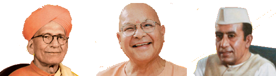
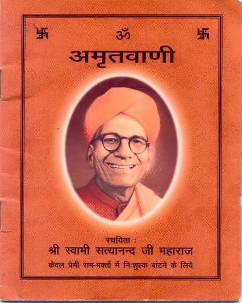 Amritwani
-
 Bhajans
-
 Ramayan
-
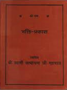 Bhakti Prakash
- Dhun
- Search
- New Clips
- Top Hits
- Catogeries
- Artists